Blank Diagram Of A Long Bone / Humerus Bone Quiz Anterior Markings - Your drawing should be in pencil.. Learn about long bone diagram with free interactive flashcards. They are one of five types of bones: Bone long blood diaphysis vector anatomical anatomy articular biology body calcium cartilage cell compact detail diagram education educational endosteum epiphysis forelimb health healthy human humerus illustration joint long bone marrow medical medicine organ orthopedic. Cheek bone (zygoma) upper jaw (maxilla). Long bones have greater length than width and consist of a shaft and a variable number of endings (extremities).
In this video we discuss the structure of bone tissue and the components of bones. Long bones follow the process of endochondral ossification where the diaphysis grows inside of cartilage from a primary ossification center until it forms most of the bone. Yours is such a clear and understandable image! A long bone has • terminal portions of the bone with thinner cortices which consist largely of cancellous bone— these are the epihyseal regions forming the articulating parts of the diaphyseal bone is organized to create the best balance between weight and structural strength. If it isn't present in your bone, draw a diagram in the blank box below to show the usual location of it.
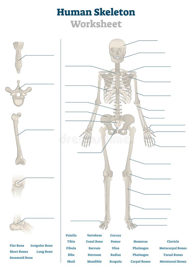
Body Movements Class 6 Extra Questions Science Chapter 8 Learn Cbse from live.staticflickr.com We make our own lab manual and need a labeled image of a human skeleton. Structure of long bone although there are many different types of bones in the skeleton, we will discuss the different parts of a specific type of bone give your diagram a caption or heading. Long, short, flat, irregular and sesamoid. If it isn't present in your bone, draw a diagram in the blank box below to show the usual location of it. During the course of development, the bone tissue is recycled, gradually altering its shape. (a) anterior view with longitudinal section spongy bone proximal epiphysis articular cartilage epiphyseal line periosteum compact bone medullary cavity diaphysis distal epiphysis (a). Related posts of diagram of of a long bone. Bone marrow (see diagram below).
Cheek bone (zygoma) upper jaw (maxilla).
Human anatomy for muscle reproductive and skeleton. Long bones follow the process of endochondral ossification where the diaphysis grows inside of cartilage from a primary ossification center until it forms most of the bone. Cheek bone (zygoma) upper jaw (maxilla). Bone marrow (see diagram below). Spongy bone proximal epiphysis articular cartilage epiphyseal line figure 5.2a the structure of a long bone (humerus). Download scientific diagram | 1 structure and components of long bone. They are usually somewhat curved under normal circumstances bones stop growing when the owner reaches his or her late teens or early twenties. During the course of development, the bone tissue is recycled, gradually altering its shape. While their parts are similar in general, their structure has. Bone marrow is the soft, highly vascular and flexible connective tissue within bone cavities which serve as the primary site of new blood cell production or bone marrow is the primary source of pluripotent stem cells that give rise to all hemopoietic cells (blood cells) including lymphocytes. The common name of each bone is listed first, with the scientific name given in parenthesis. Long bone diagram unlabled manual e books. (a) anterior view with longitudinal section spongy bone proximal epiphysis articular cartilage epiphyseal line periosteum compact bone medullary cavity diaphysis distal epiphysis (a).
We make our own lab manual and need a labeled image of a human skeleton. Dissection of a long bone in this activity you will identify the structures of a long bone and answer the questions that follow. Layer of a long bone. Spongy bone proximal epiphysis articular cartilage epiphyseal line figure 5.2a the structure of a long bone (humerus). In long bones, chondrocytes form a template of the hyaline cartilage diaphysis.
Human Skeleton Worksheet Vector Illustration Blank Educational Bone Scheme Stock Vector Illustration Of Diagram Body 157100658 from thumbs.dreamstime.com Blank diagram of the eye eye diagram blank diagram electricity free download boss od 1 anatomy of a human female back muscle anatomy human back diagram organs anatomie. Word document with a labelled diagram of the long bone which can be used as a revision aid or starter for gcse pe. Lower jaw (mandible) collar bone. Download scientific diagram | 1 structure and components of long bone. They are one of five types of bones: Just print off and cut out. Dissection of a long bone in this activity you will identify the structures of a long bone and answer the questions that follow. Related posts of diagram of of a long bone.
They are usually somewhat curved under normal circumstances bones stop growing when the owner reaches his or her late teens or early twenties.
Long bones, as their name suggests, are considerably longer than they are wide (figure 6.2a). Word document with a labelled diagram of the long bone which can be used as a revision aid or starter for gcse pe. Dissection of a long bone in this activity you will identify the structures of a long bone and answer the questions that follow. Just print off and cut out. Learn about long bone diagram with free interactive flashcards. If it isn't present in your bone, draw a diagram in the blank box below to show the usual location of it. This diagram determines the possible causes of a specific event or problem. This diagram makes it easier for one to display many potential causes for a specific effect or problem. Sectional diagram of a long bone. The hard cortical tissue can be invaded by cells that destroy the bone, called osteoclasts, only to have new bone laid down by secondary osteoblasts. We also discuss what are osteons, what are canaliculi, what are. Choose from 500 different sets of flashcards about long bone diagram on quizlet. File axial skeleton diagram blank svg wikimedia commons.
Download scientific diagram | 1 structure and components of long bone. This diagram makes it easier for one to display many potential causes for a specific effect or problem. The classification of a long bone includes having a body that is longer than it is wide, with growth plates (epiphysis) at either end, having a hard outer surface of a compact bone and a spongy inner known a. This is called the diaphysis. Since in the given question, the structure shown shows the canals helps identify the structure as osteon and is the correct answer.
19 1 Types Of Skeletal Systems Concepts Of Biology 1st Canadian Edition from opentextbc.ca Spongy bone proximal epiphysis articular cartilage epiphyseal line figure 5.2a the structure of a long bone (humerus). This diagram determines the possible causes of a specific event or problem. This is called the diaphysis. Human anatomy for muscle reproductive and skeleton. Bones of the body anatomy skeletal system labeled diagrams of the human skeleton. If it isn't present in your bone, draw a diagram in the blank box below to show the usual location of it. Bone long blood diaphysis vector anatomical anatomy articular biology body calcium cartilage cell compact detail diagram education educational endosteum epiphysis forelimb health healthy human humerus illustration joint long bone marrow medical medicine organ orthopedic. Ends (epiphyses) at the ends of the long bone, the cortex is much thinner.
Diagram of of a long bone.
Long bones, as their name suggests, are considerably longer than they are wide (figure 6.2a). Word document with a labelled diagram of the long bone which can be used as a revision aid or starter for gcse pe. Diagram of of a long bone. The long bone diagram blank could be your desire when thinking of about bone. Find out where this is usually located and, if it is present, label it on your bone. They are one of five types of bones: Long bone diagram unlabled manual e books. There is a strong ligament passing from the the humerus and the femur are corresponding bones of the arms and legs, respectively. A long bone has • terminal portions of the bone with thinner cortices which consist largely of cancellous bone— these are the epihyseal regions forming the articulating parts of the diaphyseal bone is organized to create the best balance between weight and structural strength. 9 fishbone diagram templates to get started. Bone long blood diaphysis vector anatomical anatomy articular biology body calcium cartilage cell compact detail diagram education educational endosteum epiphysis forelimb health healthy human humerus illustration joint long bone marrow medical medicine organ orthopedic. Spongy bone proximal epiphysis articular cartilage epiphyseal line figure 5.2a the structure of a long bone (humerus). Bodytomy explains the anatomy, diagram, and function of the occipital bone.
Share this post
0 Response to "Blank Diagram Of A Long Bone / Humerus Bone Quiz Anterior Markings - Your drawing should be in pencil."


0 Response to "Blank Diagram Of A Long Bone / Humerus Bone Quiz Anterior Markings - Your drawing should be in pencil."
Post a Comment